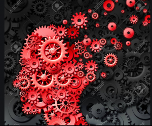
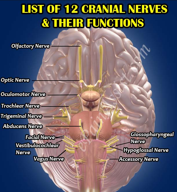
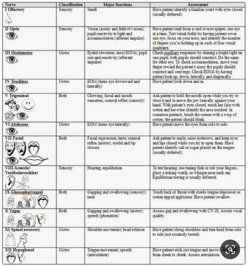
There are 12 cranial nerves, the Optic Nerve being only 1 of them. It is crucial to stress that 6 of these 12 cranial nerves innervate the eye in various degrees resulting in an elaborate neural network of visual fibres. Subsequently, over 70% of all the nerve fibres in the brain is related to visual processing.
Neuroplasticity, which is the basis of neuro-optometric rehabilitation, is the capability of these neurons & neural networks to modify their connections in response to new information from growth & development or from damage & dysfunction (concussion, brain disorders etc). Various techniques of Visual Therapy & more specifically PhotoTherapy actually induces & re-programs this neural network via sensory stimulation.
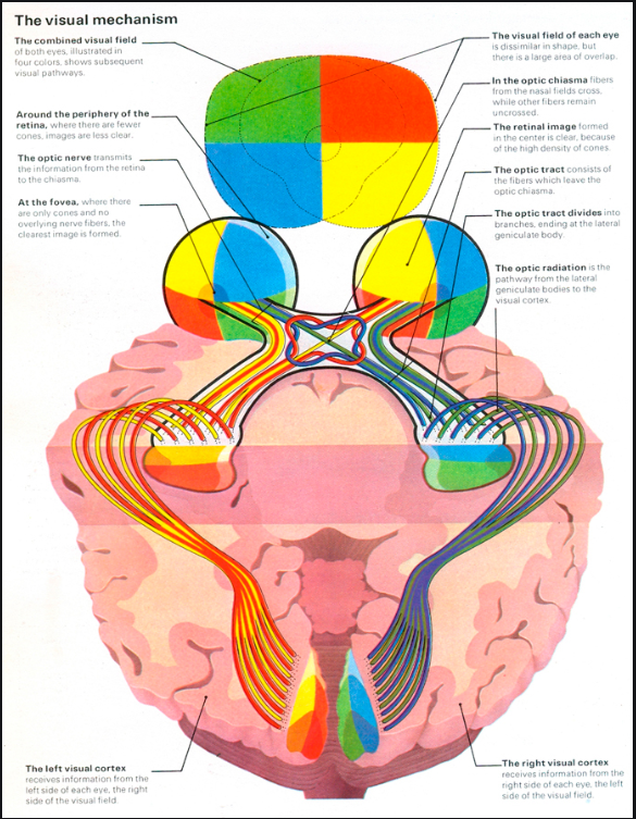
Since our visual cortex is in the back of the head (occipital lobe), the optic nerve fibers have to travel through the entire brain in a unique pattern, before we can actually perceive vision; so if any deficiency occurs anywhere along these pathways, whether developmental, disease, stress/anxiety, vascular condition or from trauma such as concussion, functional vision will be compromised to some extent - for ex. we can physically see, but we can not understand what we see!
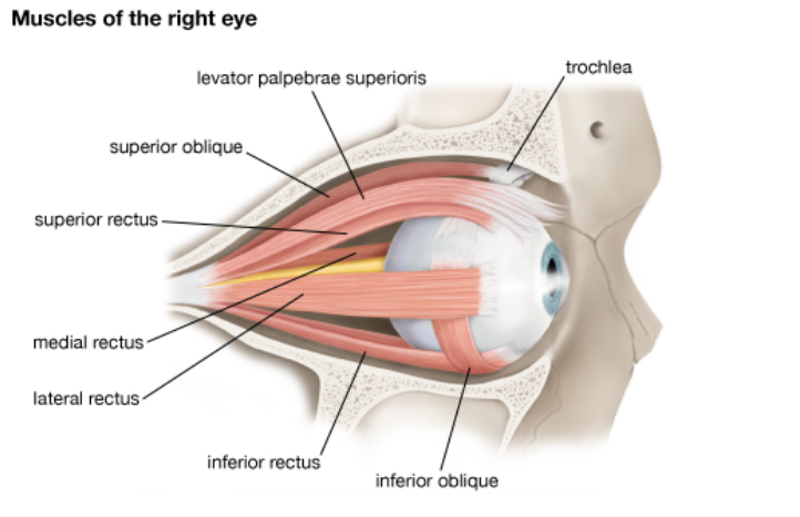
Furthermore, the muscles around the eyes (extra-ocular muscles referred to as EOM), require accurate coordination to see single & clear – comfortably.
If these muscles are unable to perform this task, symptoms such as dizziness, decreased depth perception, headaches & even double vision may result. Often kids are misdiagnosed with learning disabilities while it may have been actually an oculo-visual condition. Job performance also may manifest with inefficiencies.
If this eye mis-alignment persists, oculo-motor training ie. eye exercises may be prescribed - like physiotherapy for the eye muscles.
We utilize specialized tests to evaluate the strength of these EOMs allowing us to prescribe a personalized therapy program. Click on the tabs below.
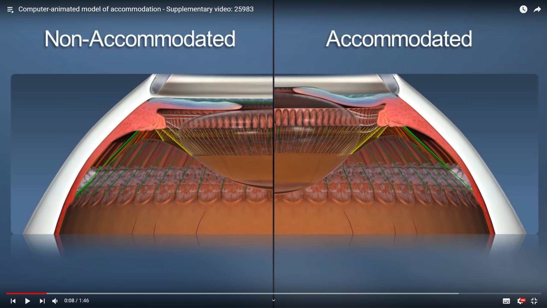
ACCOMODATION is the ability of the lens inside the eye to focus on detail at various distances. Without this lens & the ciliary muscles responsible for this focussing - we would need glasses to focus progressively at varying positions. If these ciliary muscles are weak, visual therapy eye exercises may be prescribed to strengthen them. During middle age, accomodation naturally decreases as an aging process requiring multifocals.
Vision Therapy involves training the EOMs, the nerves, visual pathways & brain centers to maximize this visual perception. The techniques used at our clinic includes the most advanced forms of therapy such as
To get a better understanding of these modalities,
you may CLICK on the above terms, or CLICK on the short video LINKS below
The Vision Therapy that we provide comprises of the following sub-specialties:
PEDIATRIC BINOCULAR VISION CONDIITONS (turned eye, lazy eye)
Pediatric Visual Therapy involves mainly the treatment of amblyopia (lazy eye), strabismus (turned eye) & strengthening the focussing muscles. Although strabismus generally includes therapy on the eye muscles, amblyopia is based more on sensory stimulation & hence the term "Neurostimulation". Newer interactive computer based techniques using the traditional patch on the dominant eye, forces the brain to interpret stimuli that is being received by the lazy eye. New cortical connections within the brain form, and the previously under or un-developed cells designated for visual perception are stimulated, leading to better resolution of visual detail. Binocular Vision is essential for learning; In 2013, published data showed 14 out of 15 oculo-motor skills were impaired in kids with reading challenges, while they achieved 20/20 on routine vision screening.
CONCUSSION / TBI (Traumatic Brain Injury)
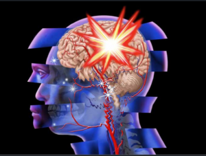
TBI such as concussions, has become a silent epidemic. Estimates indicate that TBI occurs once every 21 seconds, but due to the hidden nature of vision impairment, such as headaches, dizziness, nauzea (often assumed to be vestibular only), Ocular involvement is generally ignored despite the key role that vision plays in the brain. For example, if the eyes are 'knocked' out of alignment, then depth perception will obviously be altered, which ultimately causes these exact symptoms; since the EOMs surrounding the eye are likely to be injured during a 'hit' & over 70% of the brain involves neuro-visual processing, also affected by a 'hit', it is crucial to include Oculo-Visual Rehabilitation within the Concussion Therapy Protocols.
NEURO-OPTOMETRIC REHAB (incl VF loss from strokes etc)
ABI (Acquired Brain Injury) includes damage from medical conditions such as Stroke, Dementia, Parkinsons, Alzheimer's, MS, tumors etc. (& even concussions). Most of these conditions result in Peripheral Vision loss to some degree, along with mild overlapping of TBI symptoms. Visual-Perceptual Therapy, and neural stimulation techniques are very effective in improving the quality of Visual comfort in these conditions also.
